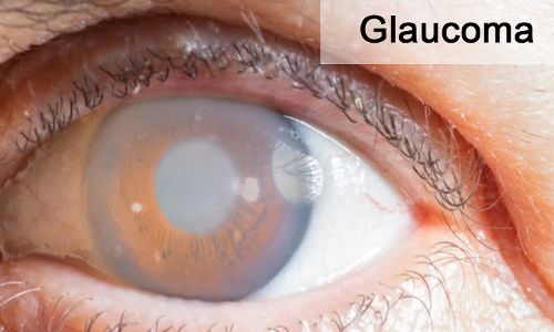
Glaucoma is a progressive optic neuropathy characterized by
structural changes in the optic nerve head and loss of the retinal nerve fiber
layer (RNFL). Elevated intraocular pressure (IOP) is the only clinically
modifiable risk factor for the progression of open-angle glaucoma. The specific
role that IOP plays in glaucomatous progression is yet to be elucidated, as
open-angle glaucoma has been found to develop across a wide spectrum of IOPs.
It is well known that IOP fluctuates throughout the day and over time and prior
research suggests that IOP variability may be a risk factor for glaucomatous
progression possibly more than IOP magnitude. However, there have been
discrepant definitions of IOP variability.
Depending on the duration of IOP monitoring, IOP
fluctuations have been measured as instantaneous, circadian
(diurnal-nocturnal), short term, and long term. Long term IOP variability can
be obtained from repeated IOP measurements taken at serial office visits over
months to years. Findings from prior studies have been inconclusive based on
assessment of the association between long-term IOP variability and visual
field (VF) progression using subjective assessment of standard automated
perimetry and/or photograph based optic disc changes.
In this study, Takashi Nishida and team evaluated the
association between longterm IOP variability and optical coherence tomography
(OCT)– based rate of RNFL thinning, an objective measure that quantifies the
retinal structural damage in patients with glaucoma. Authors also investigated
IOP variability, mean IOP, and peak IOP to compare each parameter’s association
with the rate of RNFL thinning.
In this retrospective analysis of a longitudinal cohort,
patients were enrolled from the Diagnostic Innovations in Glaucoma Study and
the African Descent and Glaucoma Evaluation study. A total of 815 eyes (564
with perimetric glaucoma and 251 with preperimetric glaucoma) from 508 patients
with imaging follow-up for a mean of 6.3 years from December 2008 to October
2020 were studied. Data were analyzed from November 2021 to March 2022.
In this study, eyes with at least 4 visits and 2 years of
follow-up optical coherence tomography and intraocular pressure measurement
were included. A linear mixed-effect model was used to investigate the
association of intraocular pressure parameters with the rates of retinal nerve
fiber layer thinning. Dominance analysis was performed to determine the
relative importance of the intraocular pressure parameters.
Of 508 included patients, 280 (55.1%) were female, 195
(38.4%) were African American, 24 (4.7%) were Asian, 281 (55.3%) were White,
and 8 (1.6%) were another race or ethnicity; the mean (SD) age was 65.5 (11.0)
years.
The mean rate of retinal nerve fiber layer change was −0.67
μm per year.
In multivariable models adjusted for mean intraocular
pressure and other confounding factors, faster annual rate of retinal nerve
fiber layer thinning was associated with a higher SD of intraocular pressure
(−0.20; P < .001) or higher intraocular pressure range (−0.05; P < .001).
In this study, higher IOP variability was independently
associated with RNFL thinning rate in patients with glaucoma. This finding is
significant, as the current mainstay of glaucoma care is lowering IOP to prevent
glaucomatous progression. This study suggests that in addition to monitoring
IOP magnitude, clinicians should consider IOP variability in the evaluation of
patients with glaucoma. In addition, this study is unique in that we estimated
the magnitude of the increase in variability explained by adding IOP
fluctuation to the RNFL slope models.
The present study helps to clarify the role of IOP
variability in both preperimetric and perimetric glaucoma; IOP variability in
terms of IOP SD and IOP range was also assessed. Additionally, as the
association of IOP variability with VF change has been studied, our study adds
to the literature by evaluating OCT-measured RNFL thinning. Authors found that
when both mean IOP and IOP variability are higher, RNFL thinning is faster.
In this cohort study, high IOP variability was independently
associated with RNFL thinning rate in patients with glaucoma. IOP fluctuation
and IOP range were more strongly associated with RNFL thinning compared with
mean IOP in the present cohort. While the biological basis for this association
is unknown, these results support the assessment IOP variability as well as
mean IOP in patients with glaucoma to identify those at greater risk of RNFL
progression.
Source: Takashi Nishida, MD, PhD; Sasan Moghimi, MD; Aimee
C. Chang, M; JAMA Ophthalmol.
doi:10.1001/jamaophthalmol.2022.4462
